
Procedures: Instrumentation and Methods
XRF Spectrometer
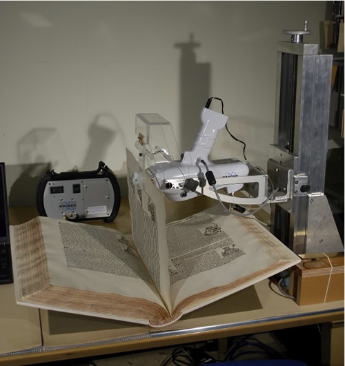
A Bruker Tracer III-V handheld XRF spectrometer fitted with a special accessory gave us data on Ca, K, S, and Fe. Jenn Wade, then at the Library of Congress’s Preservation Research and Testing Division, conducted inductively coupled plasma optical emission (ICP-OES) analyses that made possible the calibration done by Robert Shannon at Bruker Elemental. For full details on our development of the XRF instrument accessory and technique see our chapter in Current Advances in the Use of Handheld XRF in the Fields of Art Conservation and Archaeology (Leuven University Press, 2012), edited by Aaron Shugar and Jennifer L. Mass.
The abstract follows:
Historical papers vary widely in their apparent stability. Concentrations of particular elements can sometimes explain why some papers are found in browned and brittle condition and others are not. High concentrations of K, aluminum (Al), and S in such papers, for instance, are likely associated with alum, an acidic compound. Papers still durable and light in color may contain high levels of Ca, likely from the carbonate. Calcium carbonate or other compounds known to act as an alkaline reserve in paper, such as magnesium carbonate, help paper maintain an alkaline pH over time by counteracting acidic compounds that may exist within the sheet or enter it from external sources. Determination of these elemental concentrations in artifacts on paper can help the conservator make better informed decisions about the efficacy of various aqueous interventions or storage protocols.
XRF spectrometry is an especially effective nondestructive technique for elemental analysis where the density of elements of interest is high, as in the analysis of modern metals. XRF analysis of historical paper, however, is made difficult by its minimal thickness, lack of density, and the usually low concentrations and small nuclei of key elements of interest: Al, K, S, magnesium (Mg), Fe, copper (Cu), and Ca. Quantitative analysis is made more difficult by the wide variation in thickness and density of papers made in Europe between 1300 and 1800. A technique was developed to correct for the variation in paper thickness and density by placing a thin film impregnated with chromium (Cr) and bromine (Br) behind the paper specimen. Variations in Cr and Br signal strength and backscatter in a selected region of the spectrum allowed us to generate a “thickness/density” value that became part of the final calibration. A special Plexiglas accessory was constructed and mounted to a Bruker Tracer III-V handheld instrument to position the Cr/Br thin film in the same location behind paper specimens during calibration and analysis of unknowns.
Forty historical paper specimens were used in the calibration set: thirty-six to establish the calibration and four to validate the accuracy of the final calibration. Samples from ten spots were taken from each specimen and combined into one sample for destructive ICP-OES analysis. The ICP-OES data for each specimen therefore represent an average result for the ten spots. XRF analyses near each of the ten spots used for the ICP-OES sampling were collected and then summed before being used in the calibration. We assume this summed scan corresponds to the average result obtained through ICP-OES.
Using this technique, our calibration allowed us to estimate ppm concentrations of K, S, Ca, and Fe with a precision of ± 20% at a 90% confidence level when concentrations were close to 1,000 ppm. Precision improves at higher concentrations and is poorer at lower concentrations. Compositional variation must also be considered. For instance, precision is generally better with Ca, due to its high concentration. Fe was present in the calibration specimens in a narrower range, making the accuracy outside that range poorer. The precision for S is not as good as for K due to decreased instrument sensitivity for lighter elements. However, we believe this level of precision is adequate for documenting trends in the concentrations of alum (potassium aluminum sulfate or aluminum sulfate), Ca compounds, and Fe in hundreds of specimens over the centuries, or for documenting the average concentrations in large collections or collection subsets.
Our parameters for precision resulted from a review of repeated measurements made on four samples held back from the calibration. The precision reported with the results was the propagated variance and agreed with the direct measurements. The accuracy of the results on the four samples not used in the calibration, for which we had ICP-OES results, showed variations distributed fairly evenly above and below the ICP-OES values, indicating that the accuracy was much better than the precision.
We are currently evaluating results from an experiment designed to determine if this technique can be used to monitor changes in single artifacts during aqueous treatment. The precision may improve over what we obtained when analyzing widely differing unknowns because single artifacts provide the same or a very similar paper thickness/density from analysis to analysis. In the meantime, we believe the method is useful for collections surveys at its present level of development. Again, such data can provide valuable information to conservators and other preservation specialists who make collections-care and treatment decisions.
This experiment, dubbed Experiment A, employed six historical specimens, three controls, and seven aqueous treatments. XRF analyses were followed by destructive ICP-OES tests. Details on Experiment A include procedures, results, discussion, and conclusions.
UV-Vis-NIR Spectrometer
Gelatin analysis
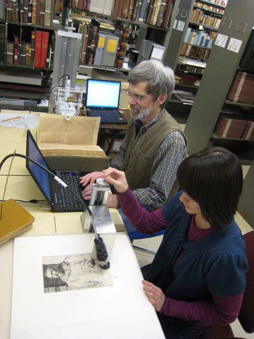
An Analytical Spectral Devices (ASD) QualitySpec Pro ultraviolet-visible-near-infrared (UV-Vis-NIR) spectrometer was used to gather data on gelatin concentration. Michael Schilling and Joy Mazurek at the Getty Conservation Institute executed the gas chromatography–mass spectrometry (GC-MS) amino acid analyses that were essential to the gelatin prediction model, which was in turn developed by Mark Ormsby at the National Archives using a similar ASD UV-Vis-NIR spectrometer. Only the NIR spectral range was used for the gelatin predictions. A manuscript detailing the instrument and technique is currently under review. A key section of the abstract follows.
Gelatin sizing was a key ingredient during the hand-papermaking era. The gelatin concentration in historical papers has never been well documented, however, because measuring the gelatin content required destructive sampling, in this project we developed a nondestructive method using near-infrared (NIR) spectrometry. Gelatin concentrations of forty historical papers dating from the fifteenth to the nineteenth century were determined from amino acid concentrations by using GC-MS. These values were combined with NIR spectra from the same papers to generate a model for predicting concentrations of unknowns. If a NIR measurement predicted a gelatin concentration in the range 0%–6%, then there was a 95% probability that the difference between the model value and a destructive amino acid measurement would be between -1.6 and +1.3 percentage points. For 6%–8% there was a 95% probability that the difference would be between -2.0 and +1.5 percentage points, and for 8%–12% the difference was between -3.0 and +2.0 percentage points.
Color analysis
Spectra gathered with this instrument also allowed us to generate a model to predict the color of each specimen. To make color measurements with the ASD instruments, which do not have the geometry specified in various color-measurement standards, working in the visible light part of the spectrum, we gathered data from the forty historical specimens used for the gelatin model calibration. At the ten gelatin sampling locations on each specimen we also took color measurements with an X-Rite spectrophotometer. By combining the readings in Excel, and with generous help from Roy Berns at the Rochester Institute of Technology’s Carlson Center for Imaging Science, we were able to fit a line to the data and determine equations for converting ASD readings into CIE L*, a*, and b* values. The R2 values were approximately 0.98. To help minimize systematic errors, we expressed our color measurements as color differences in terms of delta L*, delta a*, and delta b* values. For additional information on our color analyses see Color under DISCUSSION, Nonchronological Plots.
Strength analysis
A second experiment, dubbed Experiment B, was implemented to evaluate the ability of the UV-Vis-NIR spectrometer and an ultrasound (US) instrument (described below) to nondestructively predict the results of destructive paper-strength tests during conservation treatments. During the research, three historical specimens and two controls were tested using nondestructive UV-Vis-NIR and US methods, before and after being subjected to five different aqueous treatments common in book conservation work. Before-and-after samples from each specimen were then tested destructively. Between fifty-four and seventy-two 2 × 3 cm tabs were cut from each specimen. The historical specimen tabs were cut only from print-free areas between the chain lines, with the tabs oriented in the long direction, parallel to the chain lines. During the experiment, 342 tabs were assembled. Each received five UV-Vis-NIR and five US analyses followed by ten destructive tests using a Pulmac Zero-Span 1000 tensile tester. This totaled 3,420 nondestructive and 3,420 destructive tests. Analysis of the UV-Vis-NIR data is still underway; however, early results indicate that our UV-Vis-NIR modeling cannot predict the destructive Pulmac tests.
Experiment B details include procedures, results, discussion, and conclusions.
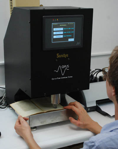
SoniSys OPUS Z-Direction Ultrasonic Tester
The ability of nondestructive ultrasonic (US) tests to predict destructive test values accurately in modern machine-made paper is well documented.1, 2, 3, 4 However, the ability of the same US tests to predict destructive test results in historical papers has never been demonstrated and was therefore a goal during this research.
Our work employed a SoniSys OPUS Z-direction ultrasonic tester that measures the rate at which sound travels through the thickness of a sheet of paper in a 1.5 cm diameter area. We first tested two 2 × 3 cm tabs from each of forty historical paper specimens using the OPUS instrument, and then followed with wet zero-span and dry short-span destructive tests. This work was followed by similar tests on a handmade hemp paper, BH, and the same handmade hemp paper sized with gelatin, BHG. Our results led us to conclude that US methods were not viable for estimating paper-strength values in widely varying historical specimens. The instrument was therefore not employed during the analysis of 1,578 specimens. However, we designed Experiment B to include US testing to determine if this instrument could be used to monitor changes in strength of the same specimen during conservation treatment. After 1,710 ultrasonic tests followed by 3,420 destructive tests, our results showed no useful correlation between the nondestructive US data and the destructive Pulmac tests. Again, Experiment B details include procedures, results, discussion, and conclusions.
[1] Gary A. Baum and Charles C. Habeger Jr., On-Line Estimates of Strength. IPC Technical Paper Series 187 (Appleton, WI: Institute of Paper Chemistry, 1986). http://hdl.handle.net/1853/2687.
[2] Charles C. Habeger and Gary A. Baum, “On-Line Measurement of Paper Mechanical Properties,” TAPPI Journal 69, no. 6 (1986): 106.
[3] K. G. Lantz and L. M. Chase, “On Line Measurement and Control of Strength Properties,” TAPPI Journal 71, no. 2 (1988): 75.
[4] M. T. Lu, “On-line Measurement of Strength Characteristics of a Moving Sheet,” TAPPI 58, no. 6 (1975): 80.